
Fig 4.
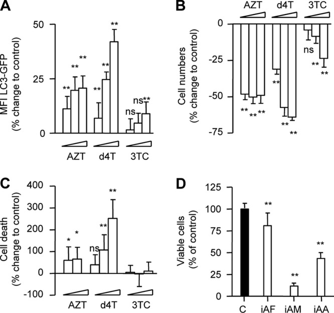
Inhibition of adipocyte autophagic flux correlates with decreased cell proliferation and viability. 3T3-F442A cells stably expressing LC3-GFP were incubated in the absence or presence of AZT (6, 30, and 150 μM), d4T (3, 15, and 75 μM), or 3TC (8, 40, and 200 μM) for 72 h. (A) Flow cytometry analysis of autophagic flux. (B) Changes in cell numbers compared to control. (C) Percentages of dead cells compared to control. (D) 3T3-F442A cells were incubated in the absence or presence of inhibitors of autophagosome formation (iAF; 3-MA [3 mM], wortmannin [30 nM], and LY294002 [7 μM]), autophagosome maturation (iAM; nocodazole [12 μM] and vinblastine [12 μM]), and autophagosome acidification (iAA; ammonium chloride [10 mM] and hydrohychloroquine and chloroquine [5 μM]) for 6 days. Data represent percentages of viable cells compared to control (= 100%). All data are presented as means ± SD and are representative of the results of at least three independent experiments with three to eight replicates. *, P < 0.05; **, P < 0.01.