
Figure 5.
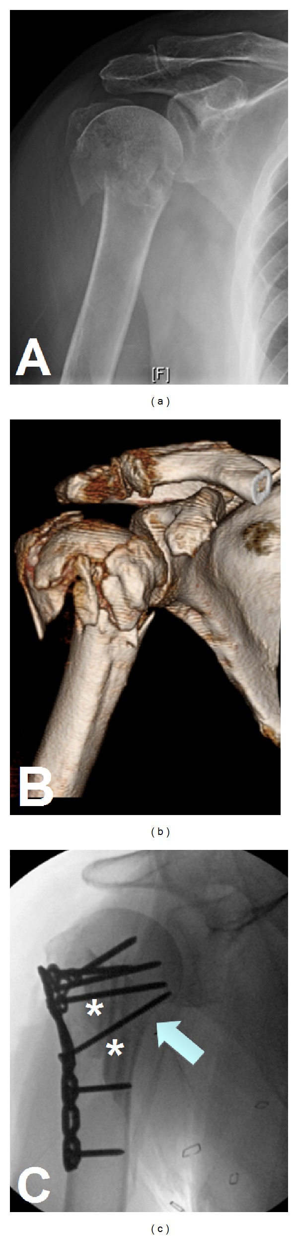
Radiograph (a) of a proximal humerus fracture. The CT scan with 3D reconstruction (b) adds significant detail and aids in preoperative planning. Osteosynthesis was carried out (c) with the use of intramedullary fibulas (asterisks) and particular attention paid to restoration of the medial calcar (arrow).