
Figure 2.
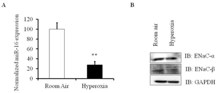
Expression levels of miR-16 and ENaC in hyperoxia induced ALI mouse models. A) miR-16 levels were analyzed by qRT-PCR in lung homogenates of mice exposed to hyperoxia and room air. Total RNA from the lung was isolated, reverse transcribed and analyzed for miR-16 using miR-16 specific assay primers and SYBR green method. miR-16 expression was normalized to 18S. B) ENaC-α and ENaC-β protein levels in mouse lung homogenates exposed to normoxia and hyperoxia were measured by western blot analysis. Lung homogenates (n=4) were subjected to immunoblotting with the indicated antibodies. **P<0.001 compared with room air controls.