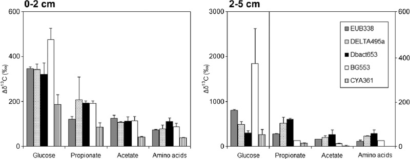
Fig 2.
The increase in δ13C ratios between labeled sediments and unlabeled control sediment (Δδ13C) for the different captured-16S rRNA fractions. Notice that the glucose data for the layer at 2 to 5 cm are plotted on a different axis (left-hand axis) than the data for the other substrates (right-hand axis), as indicated by the vertical line. The probes used in this study targeted bacteria (EUB338), Deltaproteobacteria (DELTA495a), Desulfobacteraceae (Dbact653), Gammaproteobacteria (BG553), and cyanobacteria-diatoms (CYA361). Data for 13C-amino acid-labeled sediment were normalized to the amount of 13C added with the other substrates. Part of the results determined for [13C]glucose, [13C]propionate, and [13C]acetate in the deeper layer are derived from Miyatake et al. (7); all results from the surface layer, amino acid labeling in both layers, and new target organisms (Gammaproteobacteria, cyanobacteria, and diatoms) in both layers are new data. Averages and standard deviations of the results determined for duplicate sediment incubations are presented. Captured rRNA from unlabeled controls had δ13C values between −15‰ and −20‰, within the typical range for marine phytobenthos and bacteria (23), and the δ13C values determined in duplicate analyses of unlabeled controls were within 2‰.

