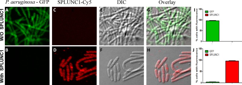
Fig 4.
Binding of SPLUNC1 to Pseudomonas aeruginosa-pMF230. Immunostaining experiments were performed without (panels A, C, E, and G) and with (panels B, D, F, and H) SPLUNC1. P. aeruginosa-pMF230 is shown in green (GFP) and SPLUNC1 in red (Cy5). (I and J) Quantitation of double fluorescence. Fluorescence was calculated using the percentages of total cells counted in 15 different randomly selected fields, and data are presented as means ± SD.

