
Fig 3.
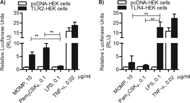
TLR2-dependent NF-κB luciferase activity. (A) TLR2-HEK cells (black bars) and pcDNA-HEK cells (white bars) incubated with MOMP proteosomes (10 μg/ml), Pam3CSK4 (0.1 μg/ml), E. coli LPS (0.1 μg/ml), or TNF-α (0.02 μg/ml) for 18 h. Luciferase activity was measured and is expressed in relative luciferase units (RLU) ± standard error normalized to nonstimulated cells (**, P = 0.002, by unpaired t test with Welch's correction; n = 6). (B) TLR4-HEK cells (black bars) and pcDNA-HEK cells (white bars) incubated as described above (**, P = 0.006 and 0.003; n = 9).