Fig 1.
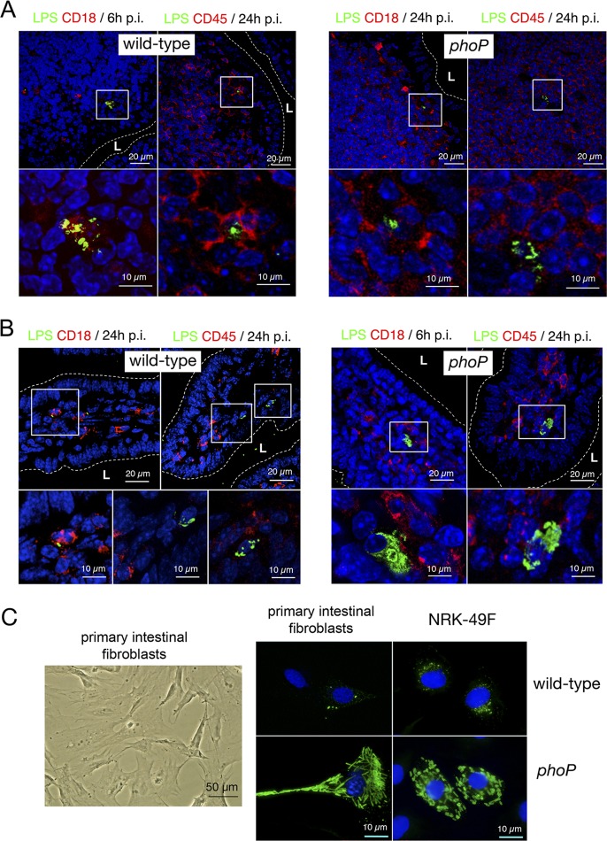
S. Typhimurium attenuates growth inside nonphagocytic cells positioned in the lamina propria of intestinal villi. (A) Tissue sections of the intestinal ileum corresponding to Peyer's patch areas were labeled with antibodies recognizing S. Typhimurium lipopolysaccharide (LPS) and the panphagocytic marker CD18 or CD45. To-pro3 was used to stain nuclei. Samples were collected at 6 or 24 h postchallenge of BALB/c mice with the SV5015 (wild-type) and MD1120 (phoP mutant) strains. Areas in boxes in upper panels are magnified in the lower panels. (B) Tissue sections showing bacterium-containing cells in the lamina propria of intestinal villi. Samples were collected at 6 or 24 h postinfection as described for panel A and were labeled with antibodies against S. Typhimurium LPS, CD18, or CD45. Note the presence of nonphagocytic stromal cells containing large numbers of intracellular phoP mutant bacteria. Areas in boxes in upper panels are magnified in the lower panels. (C) Morphology of primary intestinal fibroblasts isolated from intestinal tissue. These primary fibroblasts were infected with the SV5015 (wild-type) or MD1120 (phoP mutant) strain. In parallel, NRK-49F fibroblasts were also infected with the same strains. Bacteria were detected with anti-S. Typhimurium LPS antibodies, and nuclei were stained with 4′,6′-diamidino-2-phenylindole (DAPI). Note the similar bacterial phenotypes in both types of fibroblasts. L, intestinal lumen.

