Fig 1.
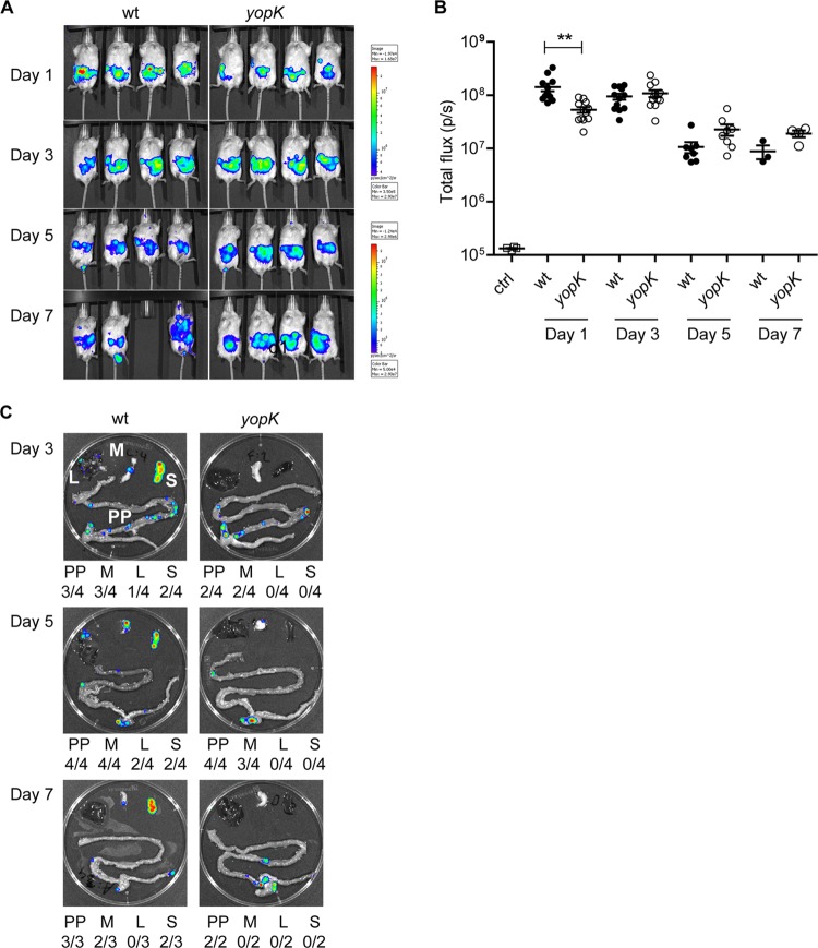
YopK is essential for Y. pseudotuberculosis to spread systemically and cause full disease in mice. Real-time in vivo imaging of Y. pseudotuberculosis infection in BALB/c mice that were orally infected with 1.1 × 10⁸ bacteria of the wild-type [YPIII(pCD1, Xen4)] or 1.3 × 10⁸ bacteria of the yopK [YPIII(pCD155, Xen4)] strains was performed. (A) Bioluminescent images of four mice for each bacterial strain on days 1, 3, 5, and 7 p.i. The same groups of mice are shown for the different time points indicated. The color bar presents the total number of emitted photons, with high and low bioluminescent signals indicated by red and blue, respectively. (B) Bioluminescent signals from the infected animals on the indicated days postinfection presented as photons/second. The data were compared by using the Student t test, with differences considered significant at a P of <0.05 (*, P < 0.05; **, P < 0.01). (C) Bioluminescent signals from a representative set of dissected organs for each bacterial strain on days 3, 5, and 7 p.i. Beneath each organ image, a summary presents the number of each indicated organ with signals/all dissected organs. PP, Peyer's patch; M, mesenteric lymph node (MLN); L, liver; S, spleen.

