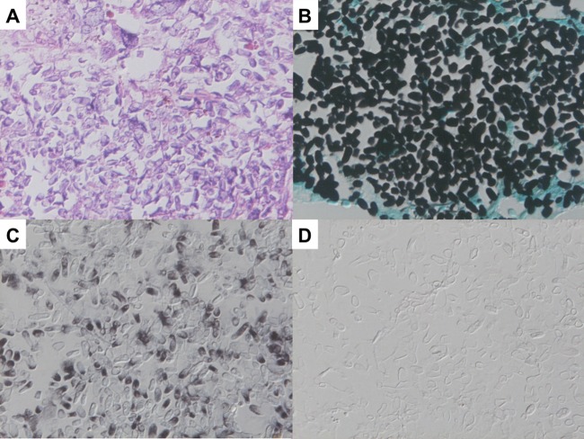
Fig 2.
Results of ISH with a pulmonary lesion of disseminated trichosporonosis confirmed by DNA sequence analysis. (A) Pathological findings with hematoxylin and eosin stain. Histological examination revealed foci consisting of yeast formations of organisms. (B) Findings with Grocott's stain. Grocott's stain showed oval or square yeast-like elements within foci of infection. (C) Result of ISH with the Trichosporon spp. PNA probe. The PNA probe against Trichosporon spp. was strongly reactive with the yeast-like elements of Trichosporon spp. (D) ISH result with the C. albicans PNA probe. The PNA probe against C. albicans was not reactive with any Trichosporon spp. organisms.

