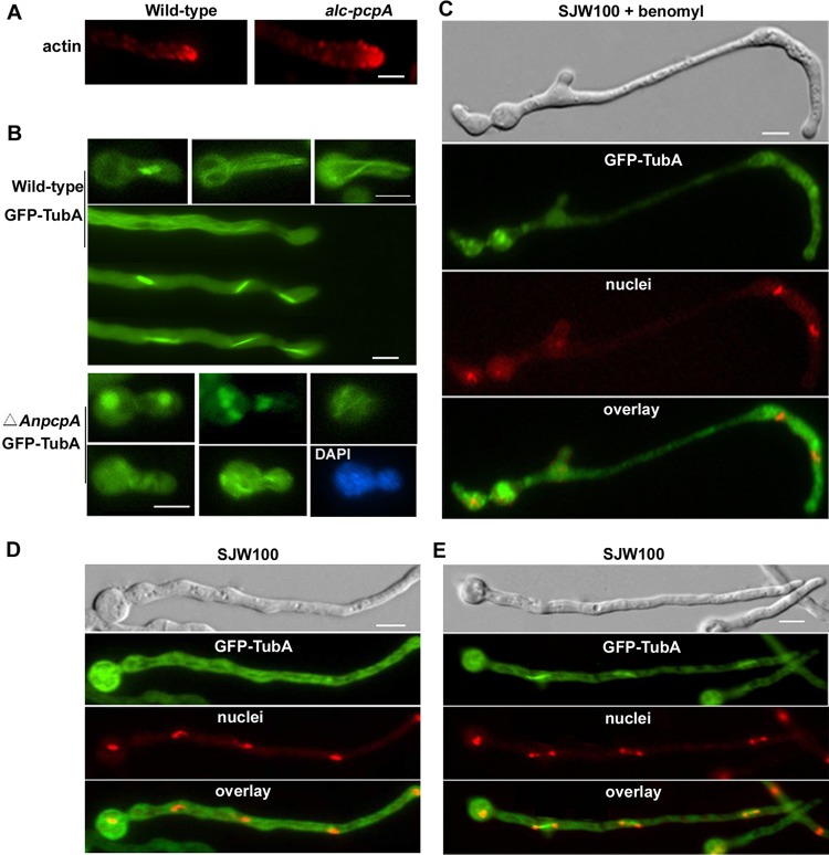
Fig 7.
Deletion of AnPcpA causes defects in microtubule organization. (A) Actin patches were visualized by indirect immunofluorescence microscopy in hyphal cells of wild-type strain WJA01 and conditional strain CPA02. (B) (Top) In control strain SJW100, GFP-TubA displayed a clear parallel bundle of interphase microtubules and mitotic spindles. (Bottom) Germlings from a strain with a deletion of AnpcpA in the SJW100 background showed very severe defects in microtubule organization and irregular GFP-TubA accumulation. Nuclei were stained with DAPI. (C) Conidial spores from SJW100 labeled with GFP-TubA were inoculated in MMGPR for 9 h and then switched to MMGPR amended with 5 μg/ml benomyl for 3 h. The nuclei were stained with DAPI. (D and E) As the control for cells shown in panel C, conidial spores from SJW100 were inoculated in MMGPR without benomyl for 12 h, the cytoplasmic microtubule was labeled with GFP-TubA, and the nuclei were stained with DAPI during interphase (D) and mitosis (E). Bars, 5 μm.

