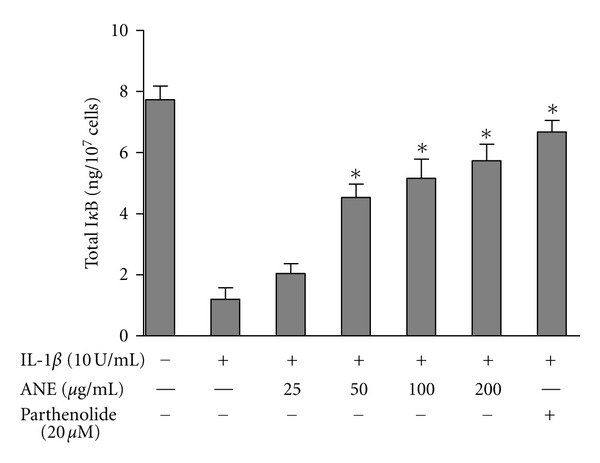
Figure 7.

Effects of BFE on IκB degradation in IL-1β-stimulated SK-N-SH cells. Cells were stimulated with IL-1β in the presence or absence of BFE (25–200 μg/mL) for 30 minutes. Cell lysates were then evaluated for IκB degradation by measuring the amount of total IκB in cell lysates. Data were analysed using one-way ANOVA for multiple comparison with post-hoc Student Newman-Keuls test. *P < 0.05 in comparison with IL-1β control.
