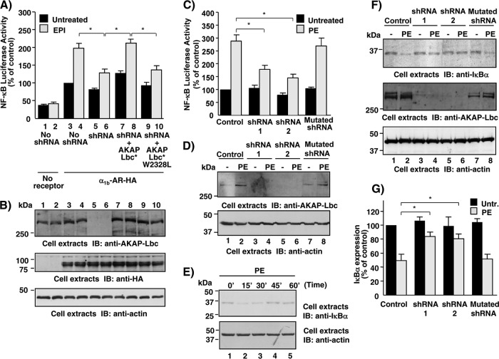
Fig 4.
AKAP-Lbc-IKKβ complex mediates α1-AR-induced NF-κB activation in cardiomyocytes. (A) HEK293 cells were infected using control lentiviruses or lentiviruses encoding AKAP-Lbc shRNAs and subsequently transfected with NF-κB–luciferase and Renilla luciferase reporter constructs in combination with either control vector (no receptor), the vector encoding the HA-tagged α1b-AR alone, or the vector encoding the HA-tagged α1b-AR together with the plasmids encoding the silencing-resistant mutants of AKAP-Lbc (AKAP-Lbc*) or AKAP-Lbc W2328L (AKAP-Lbc* W2328L). After a 24-h serum starvation, cells were treated with epinephrine (EPI) for 8 h or left untreated. Firefly luciferase activity was normalized to Renilla luciferase activity. Results are the means ± standard errors (SE) from 4 to 10 independent experiments. *, P < 0.05. (B) Expression of AKAP-Lbc, HA-α1b-AR, and actin in the lysates was assessed by Western blotting using specific antibodies as indicated. (C) Rat NVMs were infected using control lentiviruses or lentiviruses encoding wild-type or mutated AKAP-Lbc shRNAs and subsequently transfected with NF-κB–luciferase and Renilla luciferase reporter constructs. Seventy-two hours after infection, cells were incubated for 8 h in the absence or presence of 10−4 M PE. Firefly luciferase activity was normalized to Renilla luciferase activity. Results are the means ± SE from five independent experiments. *, P < 0.05. (D) Expression of AKAP-Lbc and actin in the lysates was assessed by Western blotting using specific antibodies as indicated. (E) Rat NVMs were serum starved for 24 h and subsequently treated with 10−4 M PE for the indicated periods of time. Expression of IκBα and actin in the lysates was assessed by Western blotting using specific antibodies as indicated. (F) Rat NVMs were infected as indicated in panel C. Seventy-two hours after infection, cells were incubated for 15 min in the absence or presence of 10−4 M PE. Expression of IκBα, AKAP-Lbc, and actin in the lysates was assessed by Western blotting using specific antibodies as indicated. (G) Quantitative analysis of the expression of IκBα in the cell lysates was obtained by densitometry. Untr., untreated. The amount of IκBα was normalized to the actin content of cell extracts. Results are the means ± SE from four independent experiments. *, P < 0.05.

