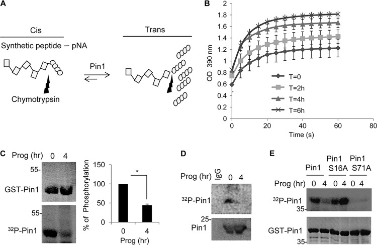
Fig 3.
Pin1 is activated during oocyte maturation. (A) Schematic to measure Pin1 activity (see the text for details). (B) Extracts from oocytes treated with progesterone for 0 to 6 h were used to assay for Pin1 activity as illustrated in panel A. (C) Extracts from oocytes treated with progesterone for 0 or 4 h were incubated with GST-Pin1 and [γ-32P]ATP; GST-Pin1 was then detected by Coomassie blue staining, and 32P-labeled GST-Pin1 was detected by autoradiography. (D) Oocytes were injected with [γ-32P]ATP, followed by progesterone treatment, immunoprecipitation of Pin1, and detection by Western blotting and autoradiography. (E) Oocytes and mature oocyte extracts were incubated with [γ-32P]ATP and GST-Pin1 or the GST-Pin1 S16A or GST-Pin1 S71A mutant proteins and analyzed by autoradiography. The lower panel shows Coomassie blue-stained GST-Pin1 isoforms.

