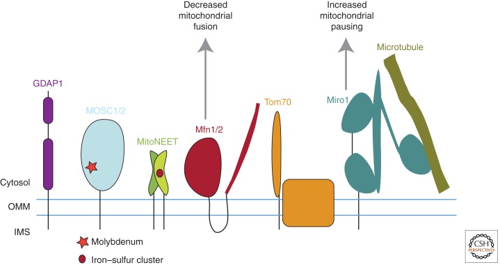
Figure 3.
Figure depicting the five mitochondrial proteins with the largest relative decrease in abundance in response to Parkin activation in unbiased quantitative proteomics experiments. They differ in the number of transmembrane domains they possess, their size, and their association with other proteins, indicating a diversity of mitochondrial substrates toward which Parkin has high activity. Three of the substrates, Mfn1, Mfn2, and Miro1, have been extensively validated as physiologic substrates of Parkin in both insects and mammals by multiple groups.

