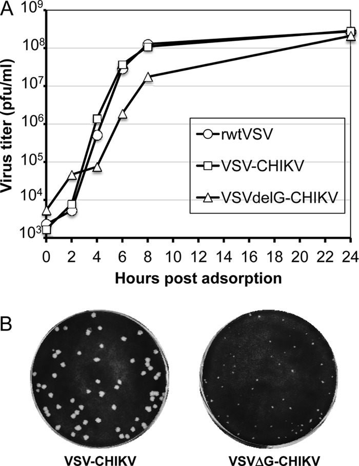
Fig 2.

Growth of recombinant VSVs and plaque morphology. (A) BHK-21 cells were infected (MOI of 10) for 30 min with rwt-VSV, VSV-CHIKV, or VSVΔG-CHIKV, and unadsorbed virus was removed by washing cells twice with PBS. Complete growth medium was added, and supernatants were collected at indicated times postadsorption. Virus titers were determined by plaque assay on BHK-21 cells. (B) Plaque morphology of VSV-CHIKV and VSVΔG-CHIKV at 2 days postinfection of BHK-21 cells.
