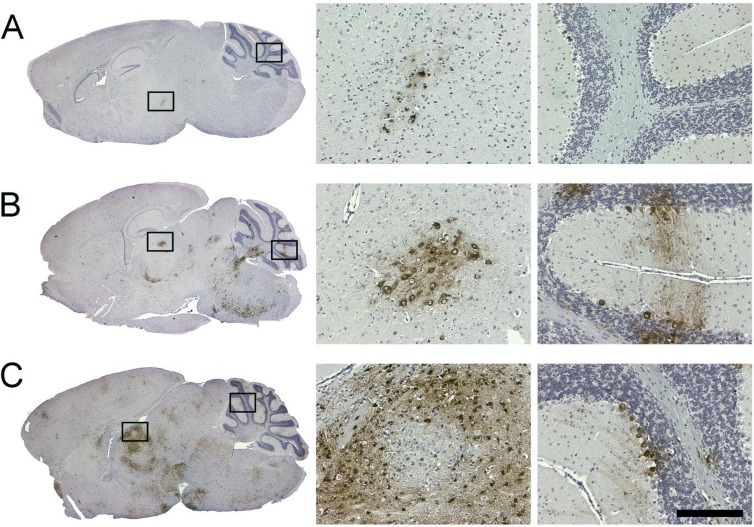
Fig 6.
Immunohistochemical analysis of mouse brain tissue collected following intraperitoneal infection. Mice infected with (A) SFV4-miRT124, (B) SFV4-miRT122, or (C) wild-type SFV4 (1 × 10⁶ PFU of virus). Samples were collected 6 days (A and B) or 5 days (C) postinfection. Seven-micrometer sagittal brain sections stained with anti-SFV antibody (left). Magnifications taken from areas indicated with black boxes (right). Scale bar, 200 μm.

