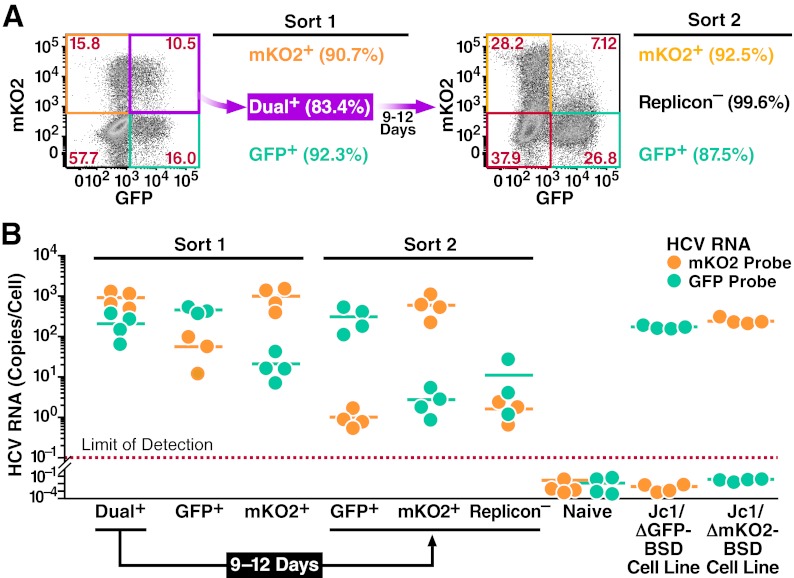
Fig 8.
Explicit loss of viral RNA during the decay process. Huh7.5 cells were transfected with Jc1/ΔGFP-BSD and Jc1/ΔmKO2-BSD RNA. Forty-eight hours later, the following populations were isolated by FACS (Sort 1): Jc1/ΔGFP-BSD-only replicon-containing cells, Jc1/ΔmKO2-BSD-only replicon-containing cells, and dual-replicon cells. The dual-replicon cells were further cultured for 9 days to allow decay to occur, and replicon-negative and single-replicon cells were isolated as before (Sort 2). The purity of each sorted population is indicated in parentheses. (A) Flow cytometric analysis of cells used for Sorts 1 and 2. Gates shown represent those used for isolation of each population. (B) Loss of viral RNA of one species during the decay process. RNA was extracted from each isolated population as well as naïve Huh7.5, stable Jc1/ΔGFP-BSD, and stable Jc1/ΔmKO2-BSD replicon-containing cell lines. Quantitative RT-PCR was used to determine the level of Jc1/ΔGFP-BSD and Jc1/ΔmKO2-BSD RNA (mKO2/GFP probes). A GAPDH probe was used for normalization of the RNA samples, using the stable Jc1/ΔGFP-BSD and Jc1/ΔmKO2-BSD cell lines as calibrators. Each data point is shown, with lines indicating the mean (n = 4 independent experiments).

