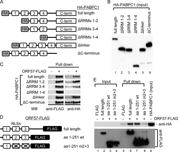
Fig 3.
Mapping the interacting regions of PABPC1 and ORF57. (A) Structures of HA-tagged wt PABPC1 and its deletion mutants. (B and C) RRMs of PABPC1 interact with ORF57. HEK293 cell lysates derived from HA-tagged wt or mt PABPC1-transfected cells (B) were incubated with FLAG only-coated (ORF57 −) or ORF57-FLAG-coated (ORF57 +) agarose beads at 4°C overnight. The IP complexes present in the pulldown assays were examined by Western blotting (WB) using anti-FLAG to recognize ORF57 and anti-HA for PABPC1 (C). (D) Structures of FLAG-tagged wt and mutant ORF57. aa, amino acids; m2+m3, mutations in both NLS2 and NLS3. (E) NLS2 and NLS3 of ORF57 interact with PABPC1. The agarose beads immobilized with wt or mt ORF57 or FLAG alone were used in the IP pulldown assays of wt PABPC1 described above, and the protein complexes present in the pulldown assays were detected with anti-HA for PABPC1 and anti-FLAG for ORF57.

