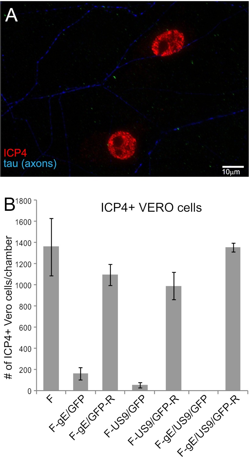
Fig 5.
Spread of gE−, US9−, and gE−/US9− mutants from distal axons to adjacent nonneuronal cells. SCG neurons were plated into microfluidic chambers, and axons were allowed to grow into the axonal compartment. Vero cells were plated in the axonal compartment 24 h before the neurons were infected with HSV-1 (8 PFU/cell) added to the somal compartments. Two to four hours after infection, 0.1% human gamma globulin was added to the axonal chambers, and 18 h after infection, the devices were disassembled. Cells in the axonal chambers were fixed with 4% paraformaldehyde, permeabilized with 0.1% Triton X-100, and immunostained with antibodies specific for the immediate-early gene product ICP4 (red) and simultaneously with antibodies specific for the axon protein tau (blue). (A) Representative image of ICP4+ Vero cells adjacent to two axons stained with tau. (B) ICP4+ Vero cells were manually counted in 10 axonal compartments involving 3 separate experiments. Total numbers of ICP4+ Vero cells per chamber are shown with standard deviations.

