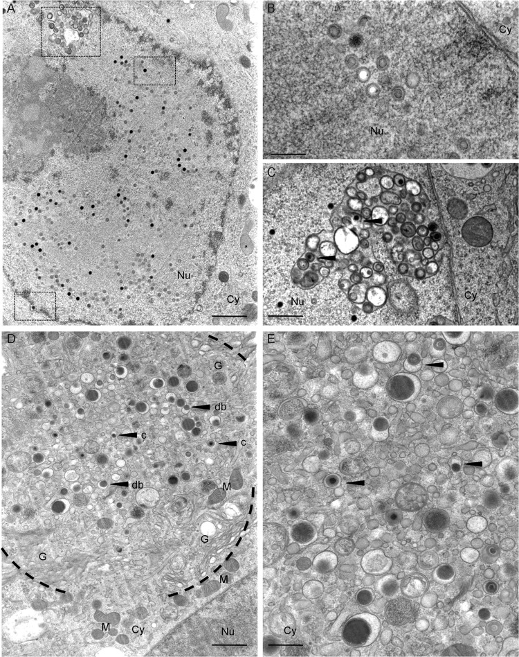
Fig 3.
Ultrastructural analysis of HCMV morphogenesis in human primary Mϕ. M2 Mϕ were infected with TB40E (MOI, 5). At 5 days postinfection, cells were fixed by high-pressure freezing, freeze-substituted, plastic embedded, and analyzed by electron microscopy after sectioning. (A) Detail of an infected nucleus showing, emphasized by the scattered squares, a cross-sectioned infolding, several scattered capsids, and a capsid budding into the perinuclear space (bar, 1 μm). (B) Magnified cross-section of the nucleus showing A (white), B (gray), and C (black) capsids (bar, 250 nm). (C) Magnified cross-section through an infolding with intermediate stages of primary envelopment visible (bar, 500 nm). (D) Overview image of an assembly compartment showing the typical rearrangement of cellular organelles and vesicles together with the accumulation of capsids (arrow c) and dense bodies (arrow db) (bar, 1 μm). (E) Magnified cross-section through the assembly compartment showing the intermediate stages of secondary envelopment (bar, 500 nm). Black arrowheads show capsids and dense bodies in close association with membranous structures. Cy, cytoplasm; Nu, nucleus; G, Golgi complex; M, mitochondria.

