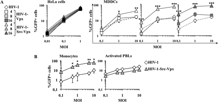
Fig 2.
HIV-1-Src-Vpx vectors display increased infectivity specifically in MDDCs and monocytes. (A) Normalized amounts of LVs (by determining their infectious titer in HeLa cells) were used at the indicated multiplicity of infection (MOI) on both HeLa cells and MDDCs. The percentage of transduced GFP-positive cells was determined 3 days later by flow cytometry in triplicate for HeLa cells and on cells from 9 donors for MDDCs. For clarity, the results obtained on MDDCs are presented on separate graphs. The results obtained upon transduction with conventional HIV-1 LVs are reported on all of the graphs for a direct comparison (dotted lines). (B) HIV-1-Src-Vpx LVs incorporating the optimal amount of Src-Vpx (16 μg) were used to transduce monocytes and PBLs that had been activated with PHA/IL-2 for 24 h prior to viral challenge. The graphs present averages and standard errors of the means (SEM) obtained with cells derived from 6 to 9 donors. Statistical analysis was performed according to an unpaired Student t test: *, P < 0.05; **, P < 0.01; ***, P < 0.001.

