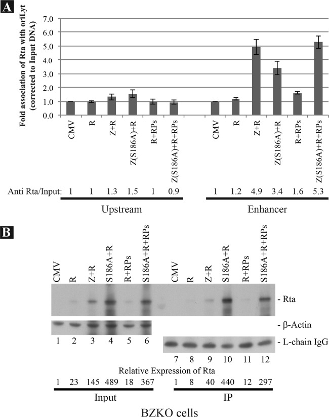
Fig 9.
wt ZEBRA and Z(S186A) promote association of Rta with the enhancer region of oriLyt. BZKO cells were transfected with expression vectors for Rta, ZEBRA, Z(S186A), or a mixture of six replication proteins (RPs) as indicated. The cells were treated with PAA at time zero and harvested at 48 h after transfection. (A) Results of a ChIP experiment analyzing the upstream and enhancer regions of oriLyt, showing immunoprecipitation with monoclonal antibody to Rta. (B) Western blot demonstrating the level of Rta protein present in the input sample and in the immunoprecipitate used for ChIP. Rabbit polyclonal anti-Rta was used to detect Rta. The ratio of cell lysate analyzed in the input versus immunoprecipitate samples was 1:7.5. β-Actin and the light chain of the anti-Rta antibody were used as loading controls for input and immunoprecipitate samples, respectively.

