Fig 2.
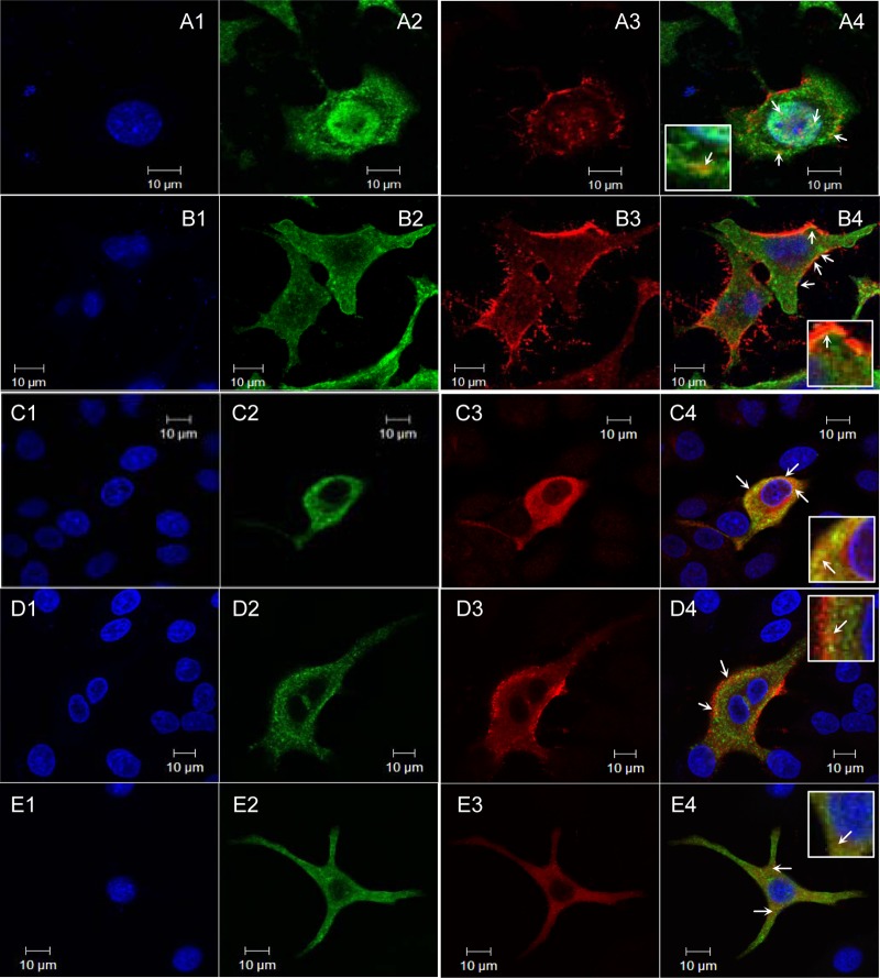
M1-NP colocalization. Following 20 h of infection (MOI, 2), MDCK cells were fixed and permeabilized before being probed with biotin-conjugated rabbit anti-M1 (red) and mouse anti-NP (green) monoclonal antibodies followed by NorthernLights streptavidin NL557 and NorthernLights 493 fluorochrome-labeled donkey anti-mouse IgG. Nucleus was counterstained by DAPI (blue). Images were acquired with a 63×, 1.4-numeric-aperture oil differential interference contrast objective lens under an LSM 510 confocal laser scanning microscope. (A1 to A4) wt-WSN; (B1 to B4) M(NLS-88R); (C1 to C4) M(NLS-88K); (D1 to D4) M(NLS-88E); (E1 to E4) M(NLS-88V). M1-NP colocalization (yellow or orange spots) is indicated by a white arrow.

