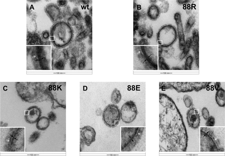
Fig 4.
Transmission electron images of M1 triple mutants. The epoxy resin-embedded M1 triple mutants were cut by an ultramicrotome, and ultrathin sections were stained with uranyl acetate and lead citrate. Images were acquired under a Zeiss EM 912 transmission electron microscope equipped with a Keenview digital camera. The inset in each transmission electron image shows an enlarged portion of the M1 layer. White arrows indicate the lipid bilayers. The inner part between two black arrows is the M1 layer. (A) wt-WSN (wt); (B) M(NLS-88R) (88R); (C) M(NLS-88K) (88K); (D) M(NLS-88E) (88E); (F) M(NLS-88V) (88V).

