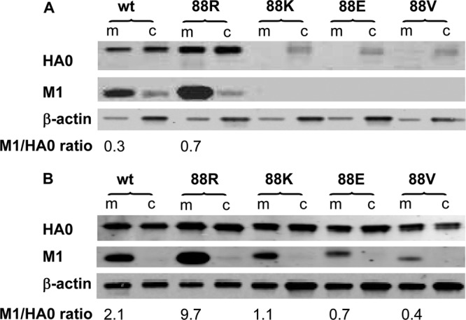
Fig 5.

Cellular membrane association of newly synthesized M1 protein. At 24 (A) or 44 (B) h postinfection (MOI, 4), MDCK cells were dissociated in hypotonic buffer, followed by low-speed centrifugation at 1,000 × g at 4°C for 10 min. The postnuclear supernatants were subjected to ultracentrifugation at 100,000 × g at 4°C for another 60 min. The pellets containing the membranes (m) were analyzed directly by Western blotting. The ultracentrifugation supernatants containing cytosol (c) were subjected to protein precipitation with ice-cold acetone before Western blotting. An equal amount of total protein was loaded per lane for each preparation of membrane and cytosol fractions. The antibody pairs included (i) sheep antiserum against HA of A/Puerto Rico/8/34 and IRDye-680LT-labeled donkey anti-sheep, (ii) biotin-conjugated rabbit anti-M1 and IRDye-680LT-labeled streptavidin, and (iii) β-actin-specific mouse monoclonal antibody and IRDye-800CW-labeled donkey anti-mouse. The blots were imaged and analyzed using an Odyssey imaging system. The M1/HA0 ratio of each virus in the corresponding membrane fraction was estimated by densitometry and is shown at the bottom of the images. wt, wt-WSN; 88R, M(NLS-88R); 88K, M(NLS-88K); 88E, M(NLS-88E); 88V, M(NLS-88V).
