
Figure 4.
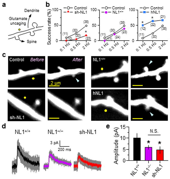
NL1 regulates activity-dependent spinogenesis
(a) Schematic of glutamate-induced spinogenesis. Dendrites of EGFP-expressing cortical layer 2/3 pyramidal neurons in acute slices from P8~12 mice were visualized with 2PLSM, and glutamate (40 pulses) was released by photolysis of caged glutamate near a low-spine density section of dendrite.
(b) Success rate of de novo spine formation in neurons where NL1 level is reduced or increased. The numbers of attempts are shown in parentheses. *:p<0.05 vs. control.
(c) Representative images of attempted spinogenesis experiments from WT neurons transfected with EGFP, sh-NL1, or hNL1, and from an EGFP-transfected NL1−/− neuron. Yellow circles and blue arrows indicate uncaging positions and, when applicable, nascent spines, respectively.
(d–e) Average dendritic NMDAR uEPSCs (d) and amplitudes (e) recorded at +40 mV in the presence of NBQX from P9-11 neurons of the indicated genotypes. Glutamate was released 0.5 μm from the dendritic shaft.