
Figure 2.
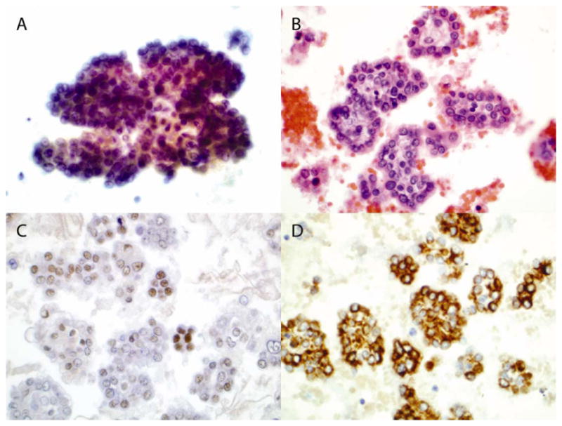
Cytology of pleural fluid (amplification, 600x). Cytospinned slide was stained with Papanicolaou stain (A). Section of cellblock was stained with H & E stain (B). Sections of cellblock were stained with antibody against WT-1 (C) and cytokeratin 5/6 (D).