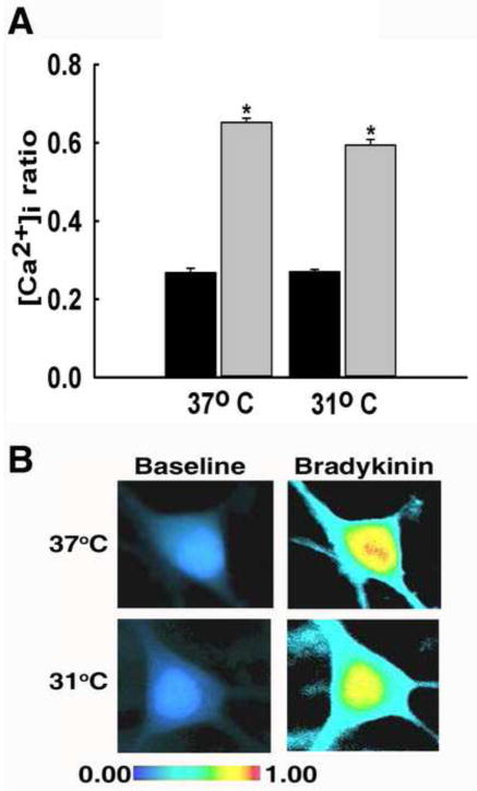
Fig. 4.
Hypothermia did not affect IP3 receptor-mediated Ca2+ entry. (A) Prior to bradykinin-mediated stimulation of IP3 receptors, neurons from 31°C and 37°C treatment groups displayed similar 340/380 baseline ratios of 0.27±0.01 and 0.27±0.01, respectively (black bars). Stimulation with 37°C bradykinin resulted in a peak 340/380 ratio of 0.65±0.03. Similarly, when stimulated with 31°C bradykinin, 340/380 ratios peaked to 0.60±0.01 (grey bars). *P<0.001 compared to 37°C baseline, one way ANOVA followed by post-hoc Tukey test, n=5 plates per condition. (B) Representative pseudocolor images obtained from baseline neuron (left panel), neuron stimulated with 37°C bradykinin (top right panel), and neuron stimulated with 31°C bradykinin (bottom right panel). Neurons in both groups exhibited elevated [Ca2+]i upon stimulation with 37°C and 31°C bradykinin.

