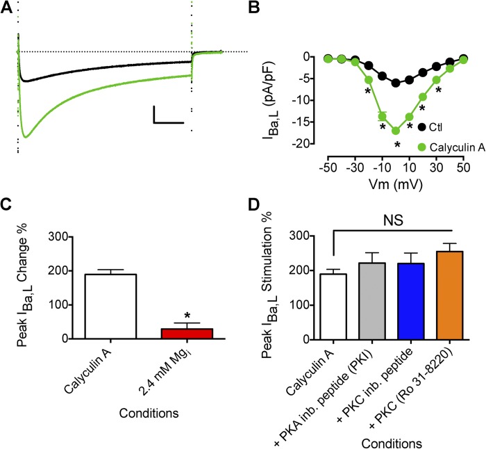
Figure 6.
Effect of Mg2+i on increased IBa,L induced by calyculin A. (A) Effect of 100 nM calyculin A on peak IBa,L (black trace is before calyculin A application). Calibration bar: 2.5 pA/pF, 50 ms. (B) Mean I-V relationship for experiments as described in A (control, n = 10; calyculin A, n = 16). (C) Effect of increased [Mg2+]i (7.2 mM; n = 6) on the increase in peak IBa,L induced by calyculin A (n = 16). (D) Effect of PKA inhibition and PKC inhibition on peak IBa,L prestimulated by calyculin A. PKA was inhibited by the preincubation of PKA inhibitor (PKI)14–22 amide, 5 µM myristoylated peptide (n = 12). PKC was inhibited by the preincubation of PKC inhibitor 20–28 amide, 24 µM myristoylated peptide (n = 8), and 1 µM Ro 31–8220 (n = 5). Myristoyl peptide inhibitors were incubated with cells for 10 min in standard extracellular solution before beginning recordings. The dotted line in A represents zero current level. Data are presented as mean ± SEM (some errors are smaller than the symbols; *, P < 0.01).

