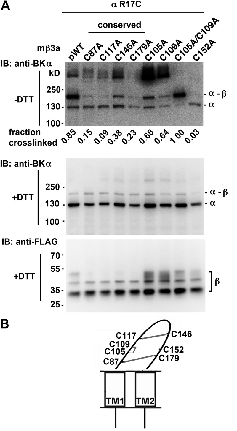
Figure 4.
Cys152 in the extracellular loop of mβ3a cross-links with α S0 R17C. (A) α S0 R17C was coexpressed with FLAG-tagged pWTβ3 or FLAG-tagged mutant β3a in which one or more of the native Cys in the extracellular loop were mutated to Ala. The intact cells were surface biotinylated, proteins were extracted, and surface proteins were purified using NeutrAvidin. The proteins were denatured in SDS and either reduced with DTT or not. The blots were developed with anti-BK α (top and middle) and anti-FLAG (bottom) antibodies. The four conserved Cys residues are indicated. Representative of more than four similar experiments. The extents of cross-linking were calculated from the relative integrated density of an ∼160-kD band divided by integrated densities of ∼160- and ∼130-kD bands. (B) Schematic of mβ3a depicting disulfide cross-linking between Cys in the extracellular loop.

