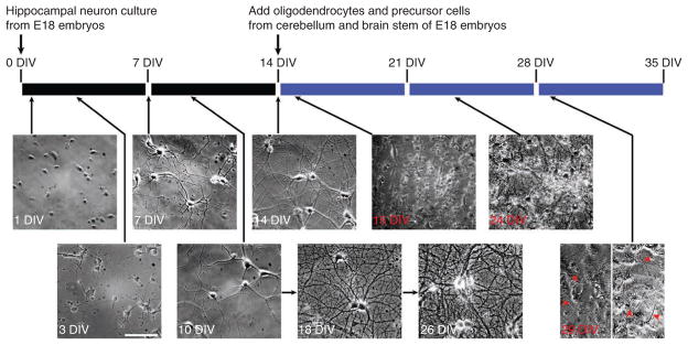
Figure 2.
The timeline for making hippocampal neuron culture and coculture. Hippocampal neurons from E18 rat embryos are dissociated and cultured for 14 d in the maintenance medium. Dividing glial cells are eliminated by the Ara-C treatment from 3 DIV to 5 DIV. Oligodendrocytes and precursor cells are dissociated from the cerebellum and brain stem of E18 rat embryos and added to the hippocampal neuron culture at 14 DIV. The maintenance medium is replaced with the myelin medium at 15 DIV. Then the culture medium is refreshed every 3 d. Myelination commences after 1–2 weeks of coculture. The example differential interference contrast microscopy images from hippocampal neuron culture (time labeled in white from 1 DIV to 26 DIV) and coculture (time labeled in red from 15 DIV to 29 DIV) at different stages are shown under the timeline. Red arrowheads, myelin segments. All images are at the same scale. Scale bar, 150 μm. Modified with permission from ref. 30. All animal experiments have been conducted in accordance with the National Institutes of Health Animal Use Guidelines and approved by the Institutional Animal Care and Use Committee at the Ohio State University.

