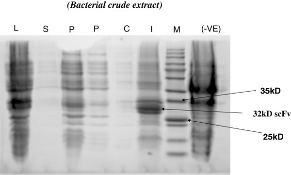
Figure 6.
Localization of scFv antibody in different fractions. Lane M, protein molecular weight marker ; lane L, cell lysate fraction; lane S, culture supernatant; lane P1 and P2 are, periplasmic fraction (20 μl and 10 μl respectively loaded); lane I, inclusion body (insoluble) fraction. Lane –Ve control, cell lysate of phagemid vector without scFv was used.

