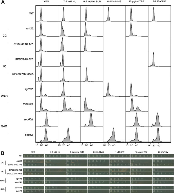
Figure 2.
Flow cytometry analysis and spot assays of eight representative mutants. (A) Flow cytometry analysis of eight mutants. Cells were grown to the logarithmic phase and treated with a DNA damage reagent for 2 h. For UV sensitivity assay, cells were exposed to 60 J/m2 radiation and then grown for 2 h. After treatment cells were harvested and subjected to cytometry analysis. (B) Sensitivity to different DNA damage reagents was quantified by spot assays. Exponentially growing cells, WT or deletions, were harvested and 5-fold serial dilutions were spotted on plates supplemented with DNA damage reagents. The plates were photographed after 3~4 days of incubation at 32°C.

