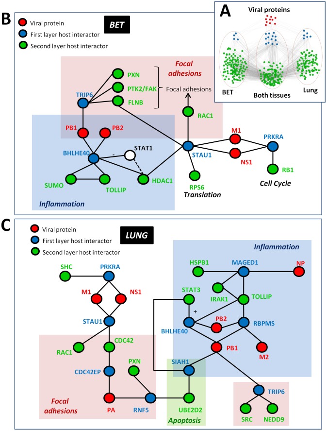
Figure 2. Tissue-specific PPI subnetwork of human proteins interacting with influenza virus proteins.
(A) Influenza proteins (red) interact with 23 ‘first layer’ host proteins (blue). These first layer proteins have interaction partners that are specific for the bronchial epithelial tissue (BET) subnetwork, for the lung subnetwork or are shared between both subnetworks (all in green). Details of the genes in this figure are given in a Cytoscape file (File S1). (B and C) Mini-networks for BET (B) and lung (C) were created from tissue-specific protein networks linking viral proteins to host proteins whose transcript was up-regulated after influenza virus infection (for a complete list of interactions, see Supplementary Table S4). Viral protein nodes are shown in red, first layer host interactors in blue and second layer host interactors in green. The STAT1 protein (shown in (B) as the white node) was not one of the original network-derived nodes, but was included due to its association with two other network nodes (BHLHE40 and HDAC1) and its known role in mediating inflammation in response to viral infection. General functions associated with different areas in each mini-network (e.g., ‘Inflammation’ and ‘Focal adhesions’) are described by partially transparent colored boxes in both (B) and (C).

