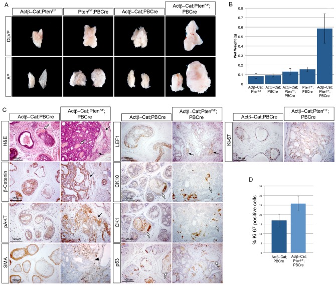
Figure 7. Stabilized β-Catenin cooperates with Pten loss to drive prostate cancer progression.
(A) brightfield images of mouse prostates with epithelial Pten deletion and stabilized β-Catenin, as indicated. The dorsal-lateral-ventral lobes (DLVP) and anterior lobes (AP) from individual animals of each genotype are shown. (B) wet weights of prostates with epithelial Pten deletion and stabilized β-Catenin. (C) H&E stain and IHC for β-Catenin, pAKT, smooth muscle actin (SMA), LEF1, CK10, CK1, p63 and Ki-67 on sections of 3-month-old Actβ-Cat;PBCre and Actβ-Cat;PtenF/F;PBCre prostates. (D) quantitative analysis of Ki-67 shows a significant increase in proliferation in Actβ-Cat;PtenF/F;PBCre prostates compared to Actβ-Cat;PBCre (p = 0.039). Black arrows indicate epithelial cells invading into the stroma. White arrows indicate areas of squamous metaplasia. Black arrowhead indicates loss of SMA. Error bars represent standard deviation.

