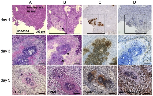
Figure 1. Histological and immunohistochemical characterization of cell infiltrates during ALA.
(A) H&E staining of mouse liver abscesses (indicated by the square in the top row of images) at the indicated times post-infection with E. histolytica trophozoites. (B) PAS staining shows E. histolytica trophozoites (arrowheads) within the abscess. (C and D) Tissue sections were stained with anti-7/4 (C) and anti-F4/80 (D) antibodies followed by HRP-conjugated secondary antibody to detect neutrophils and macrophages, respectively (brown).

