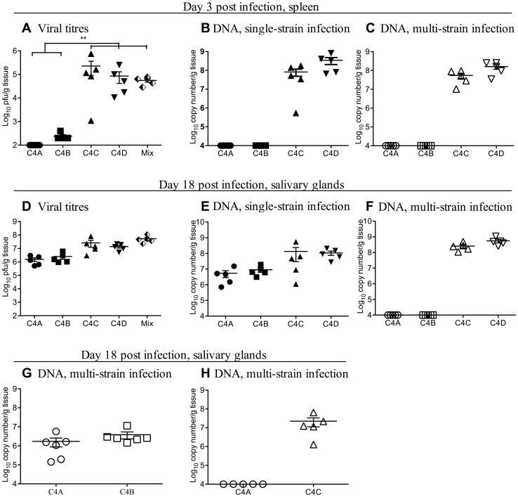
Figure 1. Competition is observed amongst co-infecting strains of MCMV in B6 mice.
The outcome of multiple MCMV infection was investigated in B6 mice during acute infection in the spleen (A–C) and persistent infection in the salivary glands (D–H). B6 mice were inoculated i.p. with 1×10⁴ pfu of either a single MCMV strain (closed symbols) or a total of 1×10⁴ pfu of an mixed inoculum of up to four MCMV strains in equal proportions (half filled diamonds - plaque assay or open symbols – strain specific PCR). A. Viral titers in the spleens of B6 mice 3 days after single strain (closed symbols) or multi-strain (Mix - half filled diamond) infection. Titers of virus in C4C infected mice were significantly higher (p<0.05) than in all other groups of MCMV infected mice. B. Viral DNA levels in the spleen of single strain infected mice from Fig. 1A. C. Viral DNA levels in the spleen of multi-strain infected mice from Fig. 1A. D. Viral titers in the salivary glands of B6 mice 18 days after single strain (closed symbols) and multi-strain infection (Mix - half filled diamond). E. Viral DNA levels in the salivary glands of single strain infected mice from Fig. 1D. Note despite low titers in the spleen, both C4A and C4B replicate in the salivary glands of B6 mice. F. Viral DNA levels in the salivary glands of multi-strain infected mice from Fig. 1D. Note during multi-strain infection both C4A and C4B were undetectable by qPCR in the salivary gland. G. B6 mice were co-infected with 1×10⁴ pfu of an equal mix of C4A and C4B and the levels of each virus assessed in the salivary glands of mice 18 days later by qPCR. Note both C4A and C4B replicate at this site when co-inoculated together. H. B6 mice were co-infected with 1×10⁴ pfu of an equal mix of C4A and C4C, and the levels of each virus assessed in the salivary glands of mice 18 days later by qPCR. Note C4A DNA was undetectable at this site in B6 mice co-infected with both C4A and C4C. The x-axis represents the limit of detection for plaque assays (100 pfu/g of tissue) and the multiplex qPCR (1×10⁴ copy number/g of tissue), n = 5 mice per group.

