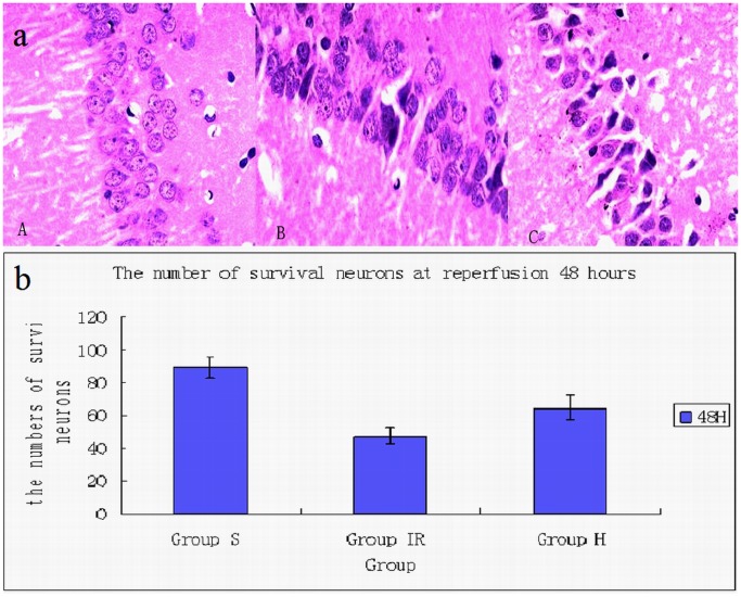
Figure 1. Neurons in hippocampus CA1 area.
(a) The picture showed the neurons in hippocampus CA1 area. The neurons in sham group (A) displayed regular appearance with large and round nuclei but pyknosis was observed in ischemia (C) and hypothermia group (B). (b) Compared with sham group(89.3±6.1) (A), the number of normal neuronal is fewer and the neurons of morphologic abnormality is more in ischemia group(47.3±4.5) (C). The number of survival neurons in hypothermia group(64.5±7.5) (B) is more than that in ischemia group. /400×visual field.

