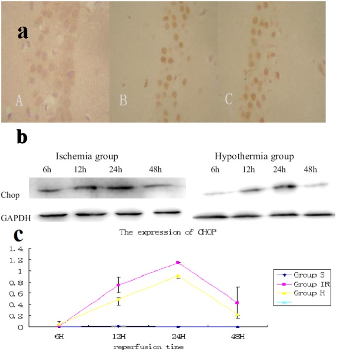
Figure 3. Expression of chop in hippocampus CA1.
(a) Immunohistochemistry showed the chop was barely detected in sham group (A).The expression of chop in hypothermia group (B) is much weaker than that in ischemia group (C) at reperfusion 24 hours. /400×visual field (b) Western blot analysis showed that the chop was barely detected in sham group. In brains of ischemia group, it was increased 6 hour after 15 minutes of ischemia and gradually decreased thereafter; however, the degree of increase was much smaller in the hypothermia brains. (c) Quantitative analysis of Western blotting showed that hypothermia after ischemia significantly decreased chop after 15 minutes of ischemia (P<0.05 compared with ischemia brains at the same time points. 6 rats from each group at every time points were used for analysis).

