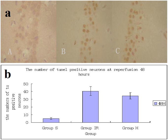
Figure 4. Neuronal apoptosis in CA1 region of hippocampus induced by global cerebral ischemia.
Detection of apoptosis in hippocampus CA1 pyramidal neurons was carried out using Tunel staing. The sham group showed a large number of neurons and almost no TUNEL-positive cells (5.1±1.2) (A). In ischemia (C) and hypothermia (B) groups, the number of neurons were decreased and substantial TUNEL-positive cells were detected. The number of TUNEL-positive cells in hypothermia group (34.4±4.2) (B) is more than in ischemia group (40.5±5.7) (C), /400×visual field.

