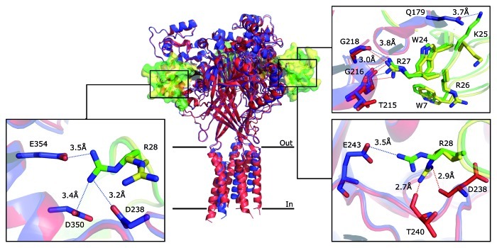
Figure 2. Molecular interactions between PcTx1 and cASIC1. The superposition of the structure obtained by Dawson et al. (PDB ID 3S3X, blue) and the low-pH structure obtained by Baconguis and Gouaux (PDB ID 4FZ0, red) is shown in cartoon representation. PcTx1 is shown in solvent-accessible surface representation (Dawson in green and Baconguis in yellow). The discrepancies of the two structures concerning their molecular interactions are illustrated in boxes; for details see text. Blue dashed lines indicate the possible hydrogen bonds in the structure obtained by Dawson et al. and red dashed lines in the structure obtained by Baconguis and Gouaux.

An official website of the United States government
Here's how you know
Official websites use .gov
A
.gov website belongs to an official
government organization in the United States.
Secure .gov websites use HTTPS
A lock (
) or https:// means you've safely
connected to the .gov website. Share sensitive
information only on official, secure websites.
