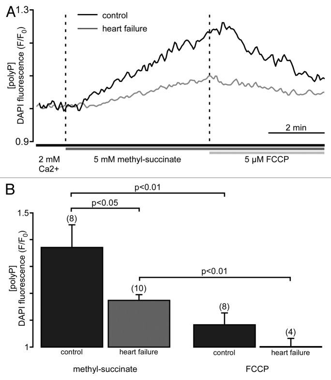
Figure 1. Mitochondrial polyP concentration is highly variable and depends on respiratory activity. (A) Original recordings of DAPI fluorescence changes in intact cardiac myocytes stimulated with 5 mM methyl-succinate followed by 5 µM FCCP from control (black) and failing myocytes (gray). DAPI fluorescence represents changes in polyP concentration. (B) Average values of maximal DAPI fluorescence after methyl-succinate (left) and minimal DAPI fluorescence during FCCP (right) addition in control (black) and heart failure (gray) cells.

An official website of the United States government
Here's how you know
Official websites use .gov
A
.gov website belongs to an official
government organization in the United States.
Secure .gov websites use HTTPS
A lock (
) or https:// means you've safely
connected to the .gov website. Share sensitive
information only on official, secure websites.
