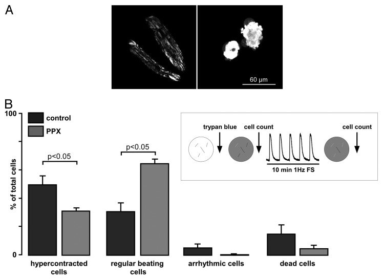
Figure 2. Polyphosphate depletion increases cell resistance to continuous work load and prevents hypercontracture. (A) Typical images of control GFP-expressing cells loaded with 10 nM tetramethylrhodamine methyl ester, a mitochondrial membrane potential sensitive dye, to point out the morphological changes seen during the stress test. Cells on the right represent an example of cells undergoing hypercontracture, which was defined as > 60% of shortening with respect to the initial cell length with concomitant morphological distortion of cell geometry (round-shaped morphology). (B) Summary of percentage of control and PPX-expressing cells (from the total amount of cells investigated), which underwent hypercontracture (left), remained normal function (regular beating cells, middle left), became arrhythmic (middle right) and died (right). Cells were stained with 0.01% Trypan blue for 10 min at room temperature.

An official website of the United States government
Here's how you know
Official websites use .gov
A
.gov website belongs to an official
government organization in the United States.
Secure .gov websites use HTTPS
A lock (
) or https:// means you've safely
connected to the .gov website. Share sensitive
information only on official, secure websites.
