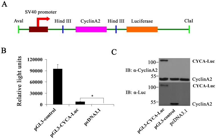
Figure 1. Luciferase activity or immunoblot analysis in U2OS cells following short-term transfection of the wild-type luciferase (Luc) and CYCA-Luc.
(A) Schematic diagrams of cyclinA2-luciferase constructs. The recombinant plasmid pGL3-CYCA-Luc encoding CYCA-Luc fusion protein contained the cyclin A2 gene fused in-frame at the N termini of the luciferase gene. (B, C) U2OS cells were placed into wells of a 6 well plate and transfected using the Lipofectamine 2000 with equal concentrations of plasmid DNA encoding either wild-type luc or CYCA-Luc chimera; pcDNA3.1 plasmid was used as a negative control. Transfected cells were cultured for 48 h, lysed, and cell extracts were assayed for luciferase activity (B), or were immunoblotted with the indicated antibodies (C). For normalization of CYCA-Luc activity or Luc, the signal (1 µg protein) for control was set to 1. This experiment was repeated three times (n = 3); error bars indicate standard error; *, p<0.05 compared with control.

