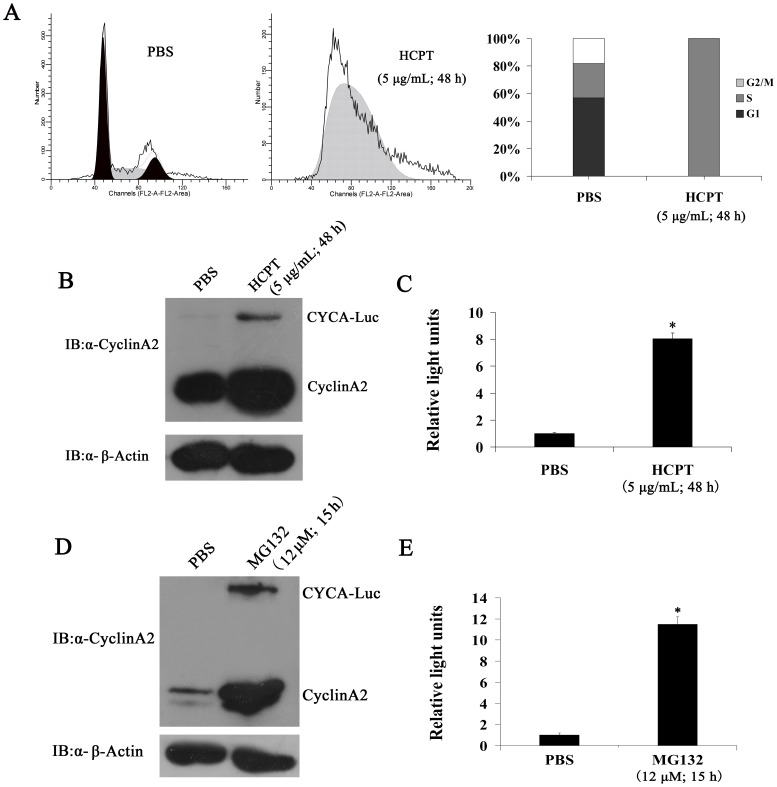
Figure 3. CYCA-Luc accumulates in response to S-phase blockage, and is degraded via the ubiquitin-proteasome pathway in HeLa-CYCA-Luc cells.
(A) After treatment with 5 µg/mL HCPT for 48 h, HeLa-CYCA-Luc cells were analyzed for DNA content by FACS after propidium iodide staining or were lysed. (B, C) Cell extracts were analyzed by immunoblotting (B) or assayed for luciferase activity (C). After treatment with 12 µmol MG132 for 15 h, HeLa-CYCA-Luc cells were lysed, and cell lysates were analyzed by immunoblotting (D) or assayed for luciferase activity (E). For normalization of luciferase or CYCA-Luc activity, the signal for untreated cells was set to 1. This experiment was repeated three times (n = 3). Error bars indicate standard error; *, p<0.05 compared with PBS.

