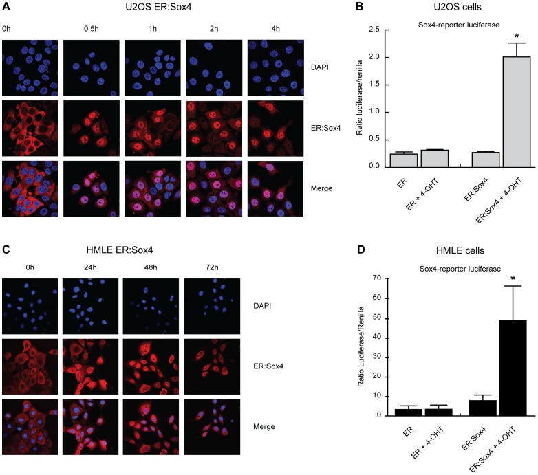
Figure 2. Generation of a Sox4 conditional activation system.
The hormone binding domain of the ER was fused to the N-terminus of Sox4. (A) ER:Sox4 was stably transduced in U2OS cells. Immuno-fluorescence analysis of ER:Sox4 localization using an anti-ER antibody after stimulation with 4-OHT (100 nM) for the time points indicated. (B) U2OS cells expressing ER and ER:Sox4 were transfected with an optimal Sox4 luciferase reporter construct and treated overnight with 4-OHT (100 nM) after which luciferase activity was measured. (C) ER:Sox4 localization in HMLE cells in presence and absence of 4-OHT. (D) HMLE cells expressing ER and ER:Sox4 were transfected with an optimal Sox4 luciferase reporter construct and stimulated overnight with 4-OHT (100 nM) after which luciferase activity was measured. Confocal microscopy data is representative of at least three independent experiments. *p<0,05 (N = 3±SD).

