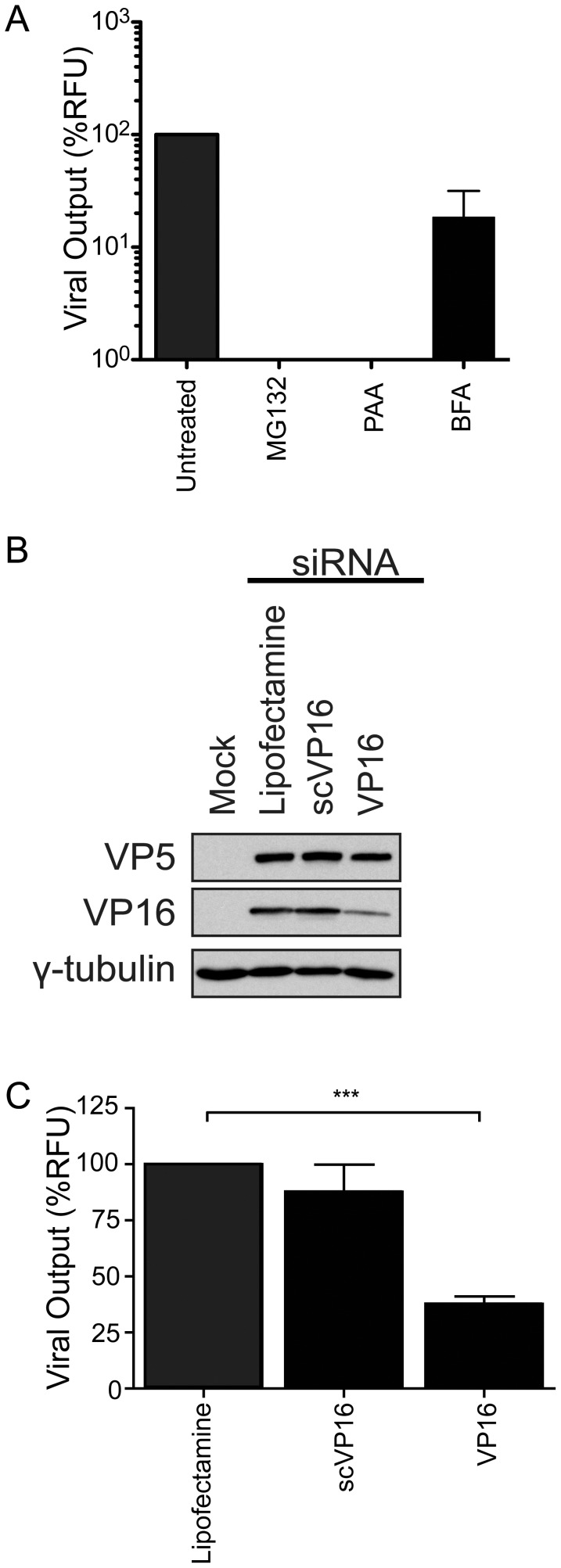
Figure 2. Validation of the assay.
143B cells were seeded in 24-well plates and infected with HSV-1 K26GFP and A) treated with drugs targeting HSV-1 post-entry (MG132), replication (PAA) and egress (BFA). B–C) As above except that cells were instead transfected for 48 hours prior to infection with Lipofectamine only, a scrambled version of the VP16 siRNA or siRNA targeting VP16. For panel A and C, the fluorescent viruses in the supernatants were concentrated by centrifugation at 24 hpi and quantified by spectrofluorometry. For panel B, 25 µg of proteins from cell lysates were tested by Western blotting for VP16 knockdown. γ-tubulin was used as loading control. The error bars show the standard errors of the mean (SEM) of three independent experiments. Bilateral Student's T-tests were performed to detect significant hits compared to the siRNA free control (***: p<0.0001).

