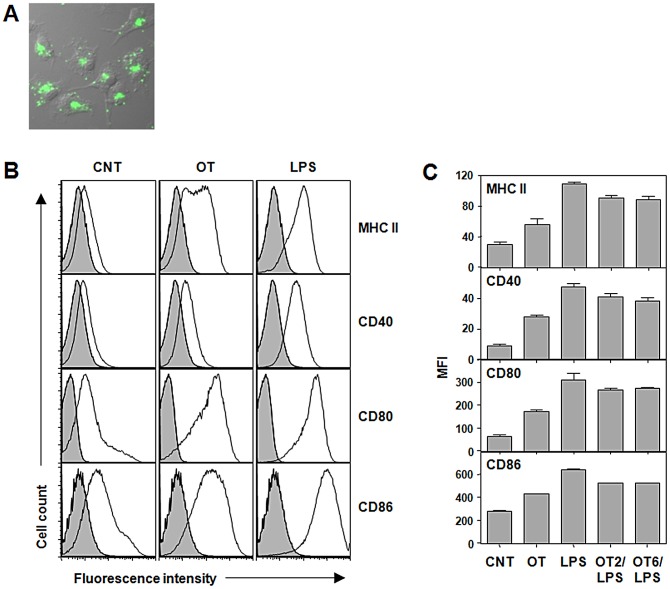
Figure 1. Activation of DCs infected with O. tsutsugamushi in vitro.
A. DCs were infected with O. tsutsugamushi for 24 h and stained with pooled scrub typhus patients' sera (green). The immunofluorescence image was merged with DIC image of the cells. B. DCs were stimulated with O. tsutsugamushi or LPS (0.5 µg/ml) for 20 h, stained with antibodies against the indicated surface molecules, and then analyzed by flow cytometer. Representative histograms of CD11c+-gated cells are presented. Gray filled: isotype control. C. The mean fluorescent intensity (MFI) of the surface markers from three separate experiments are presented. Error bar: mean+S.D., CNT: immature DCs, OT: DCs infected with O. tsutsugamushi, LPS: DCs stimulated LPS, OT2/LPS or OT6/LPS: DCs stimulated O. tsutsugamushi for 2 h (OT2) or 6 h (OT6) and then stimulated with LPS.

