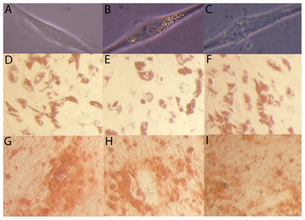
Figure 1. Morphology and differentiation potential of control and labeled adipose-derived mesenchymal stem cells.
MSCs were cultured alone or in the presence of superparamagnetic iron oxide particles (M-SPIO) or nanodiamonds for 3 days. Control MSCs (A), ∼0.9 µm M-SPIO labeled MSCs (B) and ∼0.25 µm nanodiamonds labeled MSCs (C) all exhibited similar morphologies after adhering to plastic. No significant differences were observed between the adipogenic differentiation potential of control MSCs (D), M-SPIO labeled MSCs (E) and nanodiamond labeled MSCs (F) stained with Oil Red O. Control MSCs (G), M-SPIO labeled MSCs (H) and nanodiamond labeled MSCs (I) all displayed similar levels of osteogenic differentiation following staining with Alizarin Red to visualize calcium deposition.

