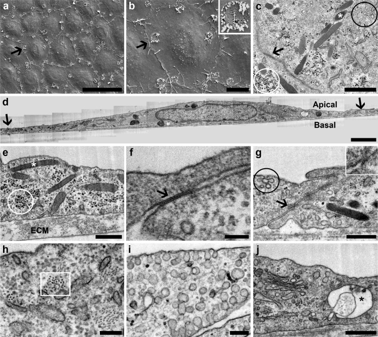
Fig. 3.
Characterization of 7-days cobblestone HUVECs by electron microscopy. HUVECs (passage 1) were seeded at 20,000 cells/cm2 on Aclar coated with Matrigel. Upon reaching the 7-days cobblestone state, cells were fixed and processed for SEM or TEM. a, b Scanning electron microscopy reveals a tight cell monolayer with well-defined cell limits. Pores (detail at inset in b) are found in the plasma membrane. Arrows point the same cell in the cobblestone. c–j Transmission electron microscopy of thin sections cut either in parallel (c) or perpendicular (d–j) to the cell monolayer shows diverse features associated to the umbilical vein endothelium in vivo; namely: Weibel-Palade bodies (c, e, white asterisks), caveolae (c, g, black circle; i), adherens junctions (f, arrow), tight junctions (g, arrow and inset), intermediate filaments (h, white square), Golgi complex (j), and glycogen inclusions (c, e, white circle). Structures resembling secretory pods, recently described for HUVECs in culture (Valentijn et al. 2010), are also present (j, black asterisk). HUVECs lie on a well-developed extracellular matrix (e, ECM). In d, a panoramic of a complete cell is shown; therein, as in c, arrows point cell limits. Scale bars: a 50 μm; b 10 μm; c 1 μm; d 2 μm; e, g, j 500 nm; f 100 nm; h, i 200 nm

